Embriões de animais evoluíram antes dos animais

quinta-feira, novembro 28, 2019

The Early Ediacaran Caveasphaera Foreshadows the Evolutionary Origin of Animal-like Embryology

Zongjun Yin6, Kelly Vargas6, John Cunningham, Maoyan Zhu, Federica Marone, Philip Donoghue

Published:November 27, 2019 DOI: https://doi.org/10.1016/j.cub.2019.10.057


Figure thumbnail fx1
Highlights

• Caveasphaera is an enigmatic component of the 609-Ma Weng’an Biota of South China

• Yin et al. use X-ray tomography to characterize cellular structure and development


• Gastrulation-like cell division, ingression, detachment, and polar aggregation occur


• A holozoan affinity suggests the early evolution of metazoan-like development

Summary

The Ediacaran Weng’an Biota (Doushantuo Formation, 609 Ma old) is a rich microfossil assemblage that preserves biological structure to a subcellular level of fidelity and encompasses a range of developmental stages [1]. However, the animal embryo interpretation of the main components of the biota has been the subject of controversy [23]. Here, we describe the development of Caveasphaera, which varies in morphology from lensoid to a hollow spheroidal cage [4] to a solid spheroid [5] but has largely evaded description and interpretation. Caveasphaera is demonstrably cellular and develops within an envelope by cell division and migration, first defining the spheroidal perimeter via anastomosing cell masses that thicken and ingress as strands of cells that detach and subsequently aggregate in a polar region. Concomitantly, the overall diameter increases as does the volume of the cell mass, but after an initial phase of reductive palinotomy, the volume of individual cells remains the same through development. The process of cell ingression, detachment, and polar aggregation is analogous to gastrulation; together with evidence of functional cell adhesion and development within an envelope, this is suggestive of a holozoan affinity. Parental investment in the embryonic development of Caveasphaera and co-occurring Tianzhushania and Spiralicellula, as well as delayed onset of later development, may reflect an adaptation to the heterogeneous nature of the early Ediacaran nearshore marine environments in which early animals evolved.


FREE PDF GRATIS: Current Biology Sup. Info.

Estrutura do gancho flagelar nativo super enrolado como junta universal: mero acaso, fortuita necessidade ou design inteligente?

quarta-feira, novembro 27, 2019

Structure of the native supercoiled flagellar hook as a universal joint

Takayuki Kato, Fumiaki Makino, Tomoko Miyata, Péter Horváth & Keiichi Namba 

Nature Communications volume 10, Article number: 5295 (2019) 

Structure of native supercoiled hook.

Abstract

The Bacterial flagellar hook is a short supercoiled tubular structure made from a helical assembly of the hook protein FlgE. The hook acts as a universal joint that connects the flagellar basal body and filament, and smoothly transmits torque generated by the rotary motor to the helical filament propeller. In peritrichously flagellated bacteria, the hook allows the filaments to form a bundle behind the cell for swimming, and for the bundle to fall apart for tumbling. Here we report a native supercoiled hook structure at 3.6 Å resolution by cryoEM single particle image analysis of the polyhook. The atomic model built into the three-dimensional (3D) density map reveals the changes in subunit conformation and intersubunit interactions that occur upon compression and extension of the 11 protofilaments during their smoke ring-like rotation. These observations reveal how the hook functions as a dynamic molecular universal joint with high bending flexibility and twisting rigidity.

O pipeline de rastreamento de células revela como os circuitos de motores são construídos: mero acaso, fortuita necessidade ou design inteligente?

terça-feira, novembro 26, 2019

Cell Volume 179, ISSUE 2, P355-372.e23, October 03, 2019

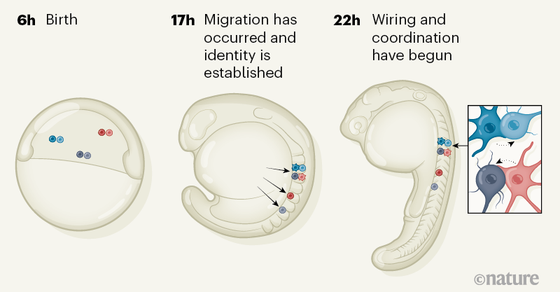
Single-Cell Reconstruction of Emerging Population Activity in an Entire Developing Circuit

Yinan Wan, Ziqiang Wei, Loren L. Looger, Minoru Koyama, Shaul Druckmann, Philipp J. Keller


Source/Fonte: Nature

Highlights

• Neurons are tracked from birth to entire circuit at cell-type and functional levels

• Neurogenesis and emergence of coordinated activity is analyzed at a single-cell level

• Motoneurons, active first, form ensembles that synchronize globally, based on size

• Neuron maturation is stereotyped, based on birth time and anatomical origin

Summary

Animal survival requires a functioning nervous system to develop during embryogenesis. Newborn neurons must assemble into circuits producing activity patterns capable of instructing behaviors. Elucidating how this process is coordinated requires new methods that follow maturation and activity of all cells across a developing circuit. We present an imaging method for comprehensively tracking neuron lineages, movements, molecular identities, and activity in the entire developing zebrafish spinal cord, from neurogenesis until the emergence of patterned activity instructing the earliest spontaneous motor behavior. We found that motoneurons are active first and form local patterned ensembles with neighboring neurons. These ensembles merge, synchronize globally after reaching a threshold size, and finally recruit commissural interneurons to orchestrate the left-right alternating patterns important for locomotion in vertebrates. Individual neurons undergo functional maturation stereotypically based on their birth time and anatomical origin. Our study provides a general strategy for reconstructing how functioning circuits emerge during embryogenesis.
 
 
FREE PDF GRATIS: Cell

Pare com o treinamento científico que exige "não pergunte"!

sexta-feira, novembro 22, 2019

WORLD VIEW 19 NOVEMBER 2019

Stop the science training that demands ‘don’t ask’

It’s time to trust students to handle doubt and diversity in science, says Jerry Ravetz.

Jerry Ravetz


As a child, I realized that my parents spoke in Yiddish when they didn’t want me to know what they were talking about, so I became aware that some knowledge was intended only for grown-ups — don’t ask. In college, I was taught an elegant theory of chemical combination based on excess electrons going into holes in the orbital shell of a neighbouring atom. But what about diatomic compounds like oxygen gas? Don’t ask; students aren’t ready to know. In physics, I learnt that Newton’s second law of motion is not an empirical, approximate relation such as Boyle’s and Hooke’s laws, and instead has a universal application; but what about the science of statics, in which forces are balanced and there is no acceleration? Don’t ask. Mere students are not worthy of an answer. Yet when I was moonlighting in the social sciences and humanities, I found my questions and opinions were respected, even if only as part of my learning experience.

Observant students will notice that social problems surrounding science are seldom mentioned in official curricula. And now, these pupils are starting to act. They have shamed their seniors into including more diverse contributors as faculty members and role models. Young scholars insolently ask their superiors why they fail to address the extinction crises elucidated by their research. Such subversions are reminiscent of the mass-produced heretical pamphlets circulated by Martin Luther’s supporters at the start of the Protestant Reformation in sixteenth-century Europe. The inherited authoritarian political structures of science education are becoming brittle — but still remain largely unchanged from my own school days.

The philosopher Thomas Kuhn once compared taught science to orthodox theology. A narrow, rigid education does not prepare anyone for the complexities of scientific research, applications and policy. If we discourage students from inquiring into the real nature of scientific truths, or exploring how society shapes the questions that researchers ask, how can we prepare them to maintain public trust in science in our ‘post-truth’ world? Diversity and doubt produce creativity; we must make room for them, and stop funnelling future scientists into narrow specialties that value technique over thought.

In the 1990s, Silvio Funtowicz, a philosopher of science, and I developed the concept of ‘post-normal science’, building on the Kuhnian terms ‘normal’ and ‘revolutionary’ science. It outlines how to use science in a society confronted with high-stakes decisions, where both facts and values are uncertain; it requires drawing on a broad community with broad inquiries. Suppressing questions from budding scientists is sure to suppress promising ideas and solutions.

...

FREE PDF GRATIS: Nature

Sabe aquele DNA "Lixo"? É cheio de informação como os teóricos do Design Inteligente afirmaram!

quarta-feira, novembro 13, 2019

The Genomic Code: A Pervasive Encoding/Molding of Chromatin Structures and a Solution of the “Non‐Coding DNA” Mystery

Giorgio Bernardi

First published: 08 November 2019 https://doi.org/10.1002/bies.201900106

junk-dna
Source/Fonte: Mapping Ignorance

Abstract

Recent investigations have revealed 1) that the isochores of the human genome group into two super‐families characterized by two different long‐range 3D structures, and 2) that these structures, essentially based on the distribution and topology of short sequences, mold primary chromatin domains (and define nucleosome binding). More specifically, GC‐poor, gene‐poor isochores are low‐heterogeneity sequences with oligo‐A spikes that mold the lamina‐associated domains (LADs), whereas GC‐rich, gene‐rich isochores are characterized by single or multiple GC peaks that mold the topologically associating domains (TADs). The formation of these “primary TADs” may be followed by extrusion under the action of cohesin and CTCF. Finally, the genomic code, which is responsible for the pervasive encoding and molding of primary chromatin domains (LADs and primary TADs, namely the “gene spaces”/“spatial compartments”) resolves the longstanding problems of “non‐coding DNA,” “junk DNA,” and “selfish DNA” leading to a new vision of the genome as shaped by DNA sequences.

FREE PDF GRATIS: BioEssays

Fios elétricos vivos conectam-se a tubos sem-fim para maior estabilidade da grade microbiana eletrogênica: mero acaso, fortuita necessidade ou design inteligente?

sexta-feira, novembro 08, 2019

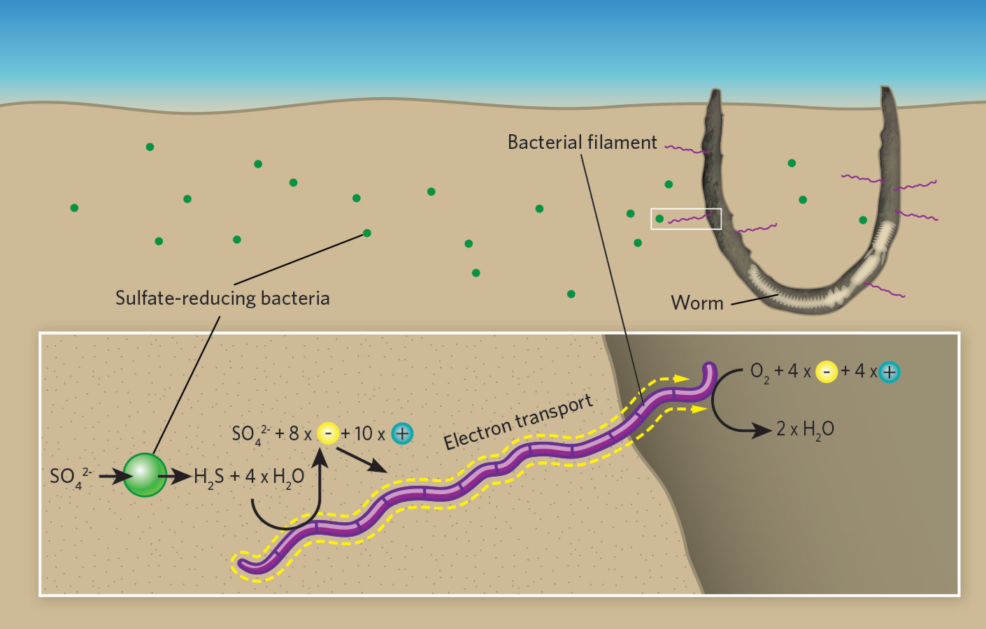
Worm tubes as conduits for the electrogenic microbial grid in marine sediments

Robert C. Aller*, Josephine Y. Aller, Qingzhi Zhu, Christina Heilbrun, Isaac Klingensmith and Aleya Kaushik†

School of Marine and Atmospheric Sciences, Stony Brook University, Stony Brook, NY 11794, USA.

↵*Corresponding author. Email: robert.aller@stonybrook.edu

↵† Present address: NOAA, Global Monitoring Division, Boulder, CO 80305, USA.

Science Advances 17 Jul 2019: Vol. 5, no. 7, eaaw3651


Abstract

Electrogenic cable bacteria can couple spatially separated redox reaction zones in marine sediments using multicellular filaments as electron conductors. Reported as generally absent from disturbed sediments, we have found subsurface cable aggregations associated with tubes of the parchment worm Chaetopterus variopedatus in otherwise intensely bioturbated deposits. Cable bacteria tap into tubes, which act as oxygenated conduits, creating a three-dimensional conducting network extending decimeters into sulfidic deposits. By elevating pH, promoting Mn, Fe-oxide precipitation in tube linings, and depleting S around tubes, they enhance tube preservation and favorable biogeochemical conditions within the tube. The presence of disseminated filaments a few cells in length away from oxygenated interfaces and the reported ability of cable bacteria to use a range of redox reaction couples suggest that these microbes are ubiquitous facultative opportunists and that long filaments are an end-member morphological adaptation to relatively stable redox domains.

FREE PDF GRATIS: Science Advances Sup. Info.

Biologia estrutural celular conforme revelada pela tomografia crioeletrônica: mero acaso, fortuita necessidade ou design inteligente?

quarta-feira, novembro 06, 2019

Cellular structural biology as revealed by cryo-electron tomography

Rossitza N. Irobalieva, Bruno Martins, Ohad Medalia

Journal of Cell Science 2016 129: 469-476; doi: 10.1242/jcs.171967


ABSTRACT

Understanding the function of cellular machines requires a thorough analysis of the structural elements that underline their function. Electron microscopy (EM) has been pivotal in providing information about cellular ultrastructure, as well as macromolecular organization. Biological materials can be physically fixed by vitrification and imaged with cryo-electron tomography (cryo-ET) in a close-to-native condition. Using this technique, one can acquire three-dimensional (3D) information about the macromolecular architecture of cells, depict unique cellular states and reconstruct molecular networks. Technical advances over the last few years, such as improved sample preparation and electron detection methods, have been instrumental in obtaining data with unprecedented structural details. This presents an exciting opportunity to explore the molecular architecture of both individual cells and multicellular organisms at nanometer to subnanometer resolution. In this Commentary, we focus on the recent developments and in situ applications of cryo-ET to cell and structural biology.

FREE PDF GRATIS: Journal of Cell Science

Faleceu Phillip E. Johnson, fundador do Movimento do Design Inteligente

sábado, novembro 02, 2019

Image result for Phillip E. Johnson
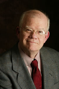
Phillip E. Johnson 1940-2019