Biologia molecular: identificando as células - mero acaso, fortuita necessidade ou design inteligente?

terça-feira, fevereiro 28, 2017

Molecular Cell - Volume 65, Issue 4, p631–643.e4, 16 February 2017

Comparative Analysis of Single-Cell RNA Sequencing Methods

Christoph Ziegenhain, Beate Vieth, Swati Parekh, Björn Reinius, Amy Guillaumet-Adkins, Martha Smets, Heinrich Leonhardt, Holger Heyn, Ines Hellmann, Wolfgang Enard7,'Correspondence information about the author Wolfgang EnardEmail the author Wolfgang Enard

7Lead Contact


Article Info

Publication History

Published: February 9, 2017 Accepted: January 17, 2017 Received in revised form: December 1, 2016 Received: August 8, 2016


Highlights

•The study represents the most comprehensive comparison of scRNA-seq protocols

•Power simulations quantify the effect of sensitivity and precision on cost efficiency

•The study offers an informed choice among six prominent scRNA-seq methods

•The study provides a framework for benchmarking future protocol improvements

Summary

Single-cell RNA sequencing (scRNA-seq) offers new possibilities to address biological and medical questions. However, systematic comparisons of the performance of diverse scRNA-seq protocols are lacking. We generated data from 583 mouse embryonic stem cells to evaluate six prominent scRNA-seq methods: CEL-seq2, Drop-seq, MARS-seq, SCRB-seq, Smart-seq, and Smart-seq2. While Smart-seq2 detected the most genes per cell and across cells, CEL-seq2, Drop-seq, MARS-seq, and SCRB-seq quantified mRNA levels with less amplification noise due to the use of unique molecular identifiers (UMIs). Power simulations at different sequencing depths showed that Drop-seq is more cost-efficient for transcriptome quantification of large numbers of cells, while MARS-seq, SCRB-seq, and Smart-seq2 are more efficient when analyzing fewer cells. Our quantitative comparison offers the basis for an informed choice among six prominent scRNA-seq methods, and it provides a framework for benchmarking further improvements of scRNA-seq protocols.

Keywords: single-cell RNA-seq, method comparison, transcriptomics, power analysis, simulation, cost-effectiveness

+++++

Professores, pesquisadores e alunos de universidades públicas e privadas com acesso ao Portal de Periódicos CAPES/MEC podem ler gratuitamente este artigo da Molecular Cell e de mais 30.000 publicações científicas. 


Dois olhos para dois propósitos: evidência in situ de visão assimétrica em lulas Histioteuthis heteropsis e Stigmatoteuthis dofleini

segunda-feira, fevereiro 27, 2017

Two eyes for two purposes: in situ evidence for asymmetric vision in the cockeyed squids Histioteuthis heteropsis and Stigmatoteuthis dofleini

Kate N. Thomas, Bruce H. Robison, Sönke Johnsen

Published 13 February 2017.DOI: 10.1098/rstb.2016.0069


Abstract

The light environment of the mesopelagic realm of the ocean changes with both depth and viewer orientation, and this has probably driven the high diversity of visual adaptations found among its inhabitants. The mesopelagic ‘cockeyed’ squids of family Histioteuthidae have unusual eyes, as the left and right eyes are dimorphic in size, shape and sometimes lens pigmentation. This dimorphism may be an adaptation to the two different sources of light in the mesopelagic realm, with the large eye oriented upward to view objects silhouetted against the dim, downwelling sunlight and the small eye oriented slightly downward to view bioluminescent point sources. We used in situ video footage from remotely operated vehicles in the Monterey Submarine Canyon to observe the orientation behaviour of 152 Histioteuthis heteropsis and nine Stigmatoteuthis dofleini. We found evidence for upward orientation in the large eye and slightly downward orientation in the small eye, which was facilitated by a tail-up oblique body orientation. We also found that 65% of adult H. heteropsis (n = 69) had yellow pigmentation in the lens of the larger left eye, which may be used to break the counterillumination camouflage of their prey. Finally, we used visual modelling to show that the visual returns provided by increasing eye size are much higher for an upward-oriented eye than for a downward-oriented eye, which may explain the development of this unique visual strategy.

This article is part of the themed issue ‘Vision in dim light’.

FREE PDF GRATIS: Phil Trans R Soc B

O tuatara é um fóssil vivo???

sexta-feira, fevereiro 24, 2017

Macroevolutionary patterns in Rhynchocephalia: is the tuatara (Sphenodon punctatus) a living fossil?

Authors

Jorge A. Herrera-Flores, Thomas L. Stubbs, Michael J. Benton

First published: 22 February 2017Full publication history


Data archiving statement Data for this study are available in the Dryad Digital Repository: https://doi.org/10.5061/dryad.568jh


Abstract

The tuatara, Sphenodon punctatus, known from 32 small islands around New Zealand, has often been noted as a classic ‘living fossil’ because of its apparently close resemblance to its Mesozoic forebears and because of a long, low-diversity history. This designation has been disputed because of the wide diversity of Mesozoic forms and because of derived adaptations in living Sphenodon. We provide a testable definition for ‘living fossils’ based on a slow rate of lineage evolution and a morphology close to the centroid of clade morphospace. We show that through their history since the Triassic, rhynchocephalians had heterogeneous rates of morphological evolution and occupied wide morphospaces during the Triassic and Jurassic, and these then declined in the Cretaceous. In particular, we demonstrate that the extant tuatara underwent unusually slow lineage evolution, and is morphologically conservative, being located near the centre of the morphospace for all Rhynchocephalia.

FREE PDF GRATIS: Palaeontology

Biologia de rede diferencial: complexidade explicada por mero acaso, fortuita necessidade ou design inteligente?

Differential network biology

Trey Ideker, Nevan J Krogan

Author Affiliations

Trey Ideker*,1,2 and Nevan J Krogan*,3,4,5

1 Departments of Medicine and Bioengineering, University of California San Diego, La Jolla, CA, USA

2 The Institute for Genomic Medicine, University of California San Diego, La Jolla, CA, USA

3 Department of Cellular and Molecular Pharmacology, University of California San Francisco, San Francisco, CA, USA

4 California Institute for Quantitative Biosciences, QB3, San Francisco, CA, USA

5 J David Gladstone Institutes, San Francisco, CA, USA

↵*Corresponding authors. Departments of Medicine and Bioengineering, University of California, 9500 Gilman Drive, San Diego, La Jolla, CA 92093, USA. Tel.: +1 858 822 4558; Fax: +1 858 822 4246; E-mail: tideker@ucsd.eduDepartment of Cellular and Molecular Pharmacology, University of California San Francisco, San Francisco, CA 94158, USA. Tel.: +1 415 476 2980; Fax: +1 415 514 9736; E-mail: krogan@cmp.ucsf.edu

DOI 10.1038/msb.2011.99 | Published online 17.01.2012

Molecular Systems Biology (2012) 8, 565

Source/Fonte: Raman Lab
Abstract

Protein and genetic interaction maps can reveal the overall physical and functional landscape of a biological system. To date, these interaction maps have typically been generated under a single condition, even though biological systems undergo differential change that is dependent on environment, tissue type, disease state, development or speciation. Several recent interaction mapping studies have demonstrated the power of differential analysis for elucidating fundamental biological responses, revealing that the architecture of an interactome can be massively re‐wired during a cellular or adaptive response. Here, we review the technological developments and experimental designs that have enabled differential network mapping at very large scales and highlight biological insight that has been derived from this type of analysis. We argue that differential network mapping, which allows for the interrogation of previously unexplored interaction spaces, will become a standard mode of network analysis in the future, just as differential gene expression and protein phosphorylation studies are already pervasive in genomic and proteomic analysis.

Genetic Interactions Networks Protein Interactions

FREE PDF GRATIS: Mol Syst Biol. 8: 565

Construção de nicho na teoria evolucionária: a construção de um nicho acadêmico???

quinta-feira, fevereiro 23, 2017

Niche construction in evolutionary theory: the construction of an academic niche?

Manan Gupta, N.G. Prasad, Sutirth Dey, Amitabh Joshi, T.N.C. Vidya


This article is a preprint and has not been peer-reviewed [what does this mean?].


Abstract

In recent years, fairly far-reaching claims have been repeatedly made about how niche construction, the modification by organisms of their environment, and that of other organisms, represents a vastly neglected phenomenon in ecological and evolutionary thought. The proponents of this view claim that the niche construction perspective greatly expands the scope of standard evolutionary theory and that niche construction deserves to be treated as a significant evolutionary process in its own right, almost at par with natural selection. Claims have also been advanced about how niche construction theory represents a substantial extension to, and re-orientation of, standard evolutionary theory, which is criticized as being narrowly gene-centric and ignoring the rich complexity and reciprocity of organism-environment interactions. We examine these claims in some detail and show that they do not stand up to scrutiny. We suggest that the manner in which niche construction theory is sought to be pushed in the literature is better viewed as an exercise in academic niche construction whereby, through incessant repetition of largely untenable claims, and the deployment of rhetorically appealing but logically dubious analogies, a receptive climate for a certain sub-discipline is sought to be manufactured within the scientific community. We see this as an unfortunate, but perhaps inevitable, nascent post-truth tendency within science.

Copyright 

The copyright holder for this preprint is the author/funder. All rights reserved. No reuse allowed without permission.

FREE PDF GRATIS: bioRxiv

O código do RNA entra em foco: mero acaso, fortuita necessidade ou design inteligente?

The RNA code comes into focus

Kelly Rae Chi

Nature 542, 503–506 (23 February 2017) doi:10.1038/542503a

Published online 22 February 2017

As researchers open up to the reality of RNA modification, an expanded epitranscriptomics toolbox takes shape.

Subject terms: Biological techniques Epigenetics Non-coding RNAs Transcriptomics

A molecular model of a bacterial ribosome bound to messenger RNA, a complex that is formed during protein synthesis. Laguna Design/SPL

INTRODUCTION

In 2004, oncologist Gideon Rechavi at Tel Aviv University in Israel and his colleagues compared all the human genomic DNA sequences then available with their corresponding messenger RNAs — the molecules that carry the information needed to make a protein from a gene. They were looking for signs that one of the nucleotide building blocks in the RNA sequence, called adenosine (A), had changed to another building block called inosine (I). This 'A-to-I editing' can alter a protein's coding sequence, and, in humans, is crucial for keeping the innate immune response in check. “It sounds simple, but in real life it was really complicated,” Rechavi recalls. “Several groups had tried it before and failed” because sequencing mistakes and single-nucleotide mutations had made the data noisy. But using a new bioinformatics approach, his team uncovered thousands of sites in the transcriptome — the complete set of mRNAs found in an organism or cell population — and later studies upped the number into the millions1.

Inosine is something of a special case: researchers can readily detect this chink in the armour by comparing DNA and RNA sequences. But at least one-quarter of our mRNAs harbour chemical tags — decorations to the A, C, G and U nucleotides — that are invisible to today's sequencing technologies. (Similar chemical tags, called epigenetic markers, are also found on DNA.) Researchers aren't sure what these chemical changes in RNA do, but they're trying to find out.

A wave of studies over the past five years — many of which focus on a specific RNA mark called N6-methyladenosine (m6A) — have mapped these alterations across transcriptomes and demonstrated their importance to health and disease. But the problem is vast: these marks coat not only mRNA but other RNA transcripts as well, and they cut across all the domains of life and beyond, marking even viruses with their presence.

The modifications themselves are not new. What has given them meaning and driven epitranscriptomics into the spotlight is the discovery of enzymes that can add, remove and interpret them. In 2010, chemical biologist Chuan He at the University of Chicago, Illinois, proposed that these chemical tags could be reversible and important regulators of gene expression. Not long afterwards, his group demonstrated2 the first eraser of these marks on mRNA, an enzyme called FTO. That discovery meant that m6A wasn't just a passive mark — cells actively controlled it. And this realization came at about the same time that global approaches, harnessing the power of next-generation sequencing, made it possible to map m6A and other modifications across the transcriptome.

...

FREE PDF GRATIS: Nature

Uma corrida em busca do ouro epigenético: novos controles para expressão gênica

An epigenetics gold rush: new controls for gene expression

How rediscovered chemical tags on DNA and RNA are shaking up the field.

Cassandra Willyard

22 February 2017


Some big ideas seem to appear out of nowhere, but in 2008 Chuan He deliberately went looking for one. The US National Institutes of Health had just launched grants to support high-risk, high-impact projects, and He, a chemist at the University of Chicago in Illinois, wanted to apply. But he needed a good pitch.

He had been studying a family of proteins that repair damaged DNA, and he began to suspect that these enzymes might also act on RNA. By a stroke of luck, he ran into molecular biologist Tao Pan, who had been investigating specific chemical marks, called methyl groups, that are present on RNAs. The pair worked in the same building at the University of Chicago, and began meeting regularly. From those conversations, their big idea took shape.

At the time, biologists were getting excited about the epigenome — the broad array of chemical marks that decorate DNA and its protein scaffold. These marks act like a chemical notation, telling the cell which genes to express and which to keep silent. As such, the epigenome helps to explain how cells with identical DNA can develop into the multitude of specialized types that make up different tissues. The marks help cells in the heart, for example, maintain their identity and not turn into neurons or fat cells. Misplaced epigenetic marks are often found in cancerous cells.

When He and Pan began working together, most epigenetic research focused on the tags associated with DNA and the histone proteins that it wraps around. But more than 100 different types of chemical mark had been identified on RNA, and nobody knew what they did. Some of the enzymes He was studying could strip off methyl groups, and He and Pan wondered whether one of them might work on RNA. If the marks could be reversed, they might constitute an entirely new way of controlling gene expression. In 2009, they got funding to hunt for reversible marks on RNA and the proteins that erase them.

Nine years later, such research has given birth to an 'ome of its own, the epitranscriptome. He and others have shown that a methyl group attached to adenine, one of the four bases in RNA, has crucial roles in cell differentiation, and may contribute to cancer, obesity and more1, 2. In 2015, He's lab and two other teams uncovered the same chemical mark on adenine bases in DNA (methyl marks had previously been found only on cytosine), suggesting that the epigenome may be even richer than previously imagined3. Research has taken off. “I think we're approaching a golden age of epigenomics and epitranscriptomics,” says Christopher Mason, a geneticist at Weill Cornell Medical College in New York City. “We can actually start to see all these modifications that we knew have been there for decades.”

...

FREE PDF GRATIS: Nature

Geração de formas de complexidade através da resolução de conflitos teciduais: mero acaso, fortuita necessidade ou design inteligente?

terça-feira, fevereiro 21, 2017

Generation of shape complexity through tissue conflict resolution

Alexandra B Rebocho Paul Southam J Richard Kennaway J Andrew Bangham Enrico Coen 

John Innes Centre, England; University of East Anglia, England


Published February 7, 2017

Cite as eLife 2017;6:e20156


Abstract

Out-of-plane tissue deformations are key morphogenetic events during plant and animal development that generate 3D shapes, such as flowers or limbs. However, the mechanisms by which spatiotemporal patterns of gene expression modify cellular behaviours to generate such deformations remain to be established. We use the Snapdragon flower as a model system to address this problem. Combining cellular analysis with tissue-level modelling, we show that an orthogonal pattern of growth orientations plays a key role in generating out-of-plane deformations. This growth pattern is most likely oriented by a polarity field, highlighted by PIN1 protein localisation, and is modulated by dorsoventral gene activity. The orthogonal growth pattern interacts with other patterns of differential growth to create tissue conflicts that shape the flower. Similar shape changes can be generated by contraction as well as growth, suggesting tissue conflict resolution provides a flexible morphogenetic mechanism for generating shape diversity in plants and animals.


eLife digest

Plant and animal organs come in many different shapes, from pitcher-shaped leaves and butterfly wings, to orchid flowers and the convoluted shape of the brain. Unlike pottery or sculpture, no external hand guides the formation of these biological structures; they arise on their own, through sheets of cells developing into particular three-dimensional shapes. But how does this process of self-making operate? We know that patterns of gene activity are important, because mutations that disrupt these patterns change the shape of the organ. But it is not clear how these patterns lead to sheets of cells curving and bending themselves into their characteristic three-dimensional shapes.

Plants are particularly useful tools for studying how three-dimensional organs form because, unlike animals, their cells do not slide relative to each other, which makes the analysis simpler. Rebocho et al. used a combination of computational modelling and cell analysis to study how the intricately shaped flowers of plants known as Snapdragons form. The experiments show that genes control the shape of Snapdragon flowers by causing groups of cells to grow at different rates and in different directions. This pattern of growth creates internal conflicts that are resolved by sheets of cells curving in particular ways, accounting for the three-dimensional shape.

Rebocho et al. propose that the principles of tissue conflict resolution described in this work may also underlie the development and evolution of many other plant and animal organ shapes. A future challenge is to identify the cellular mechanisms that link patterns of gene activity to the generation and resolution of conflicting cell behaviours.


FREE PDF GRATIS: eLIFE

Contra a Associação Brasileira Cristãos na Ciência, este livro critica o teísmo evolucionista científica, filosófica e teologicamente

segunda-feira, fevereiro 20, 2017


Theistic Evolution: A Scientific, Philosophical, and Theological Critique

Edited by J. P. Moreland, Stephen C. Meyer, Chris Shaw, Wayne Grudem

Format: Printed Caseside

Availability: Forthcoming

Expected: Nov 30, 2017

Retail Price: $55.00


About Theistic Evolution

The debate about biological origins continues to be hotly contested within the Christian church. Prominent organizations such as Biologos (USA) and Faraday Institute (UK) insist that Christians must yield to an unassailable scientific consensus in favor of contemporary evolutionary theory and modify traditional biblical ideas about the creation of life accordingly. They promote a view known as “theistic evolution” or “evolutionary creation.” They argue that God used—albeit in an undetectable way—evolutionary mechanisms to produce all forms of life. This book contests this proposal. Featuring two dozen highly credentialed scientists, philosophers, and theologians from Europe and North America, this volume provides the most comprehensive critique of theistic evolution yet produced. It documents evidential, logical, and theological problems with theistic evolution, opening the door to scientific and theological alternatives—making the book essential reading for understanding this worldview-shaping issue.

Editors


J. P. Moreland (PhD, University of Southern California) is distinguished professor of philosophy at Biola University. He is an author of, contributor to, or editor of over ninety books, including The Soul: How We Know It's Real and Why It Matters.


Stephen Meyer (PhD, University of Cambridge) is the director of the Discovery Institute's Center of Science and Culture. He is the author of several books, including the best-selling Darwin's Doubt and Signature in the Cell.


Chris Shaw (PhD, Queen's University, Belfast) is professor of drug discovery in the school of pharmacy at Queen's University in Belfast. He is the author of hundreds of peer-reviewed papers and the cofounder of a biomarker discovery company.


Wayne Grudem (PhD, University of Cambridge; DD, Westminster Theological Seminary) is research professor of theology and biblical studies at Phoenix Seminary, having previously taught for 20 years at Trinity Evangelical Divinity School. He is the former president of the Evangelical Theological Society, a member of the Translation Oversight Committee for the English Standard Version of the Bible, the general editor of the ESV Study Bible, and has published over 20 books. 


Specifications

Category: Academic

Format: Printed Caseside

Page Count: 800

ISBN-10: 1-4335-5286-8

ISBN-13: 978-1-4335-5286-1

Size: 6.0 in x 9.0 in

Weight: 24.0 ounces

Published: November 30, 2017

Darwin, os mecanismos celulares e moleculares do desenvolvimento das lentes de vertebrados: mero acaso, fortuita necessidade ou design inteligente?

The cellular and molecular mechanisms of vertebrate lens development

Aleš Cvekl, Ruth Ashery-Padan

Development 2014 141: 4432-4447; doi: 10.1242/dev.107953


Abstract

The ocular lens is a model system for understanding important aspects of embryonic development, such as cell specification and the spatiotemporally controlled formation of a three-dimensional structure. The lens, which is characterized by transparency, refraction and elasticity, is composed of a bulk mass of fiber cells attached to a sheet of lens epithelium. Although lens induction has been studied for over 100 years, recent findings have revealed a myriad of extracellular signaling pathways and gene regulatory networks, integrated and executed by the transcription factor Pax6, that are required for lens formation in vertebrates. This Review summarizes recent progress in the field, emphasizing the interplay between the diverse regulatory mechanisms employed to form lens progenitor and precursor cells and highlighting novel opportunities to fill gaps in our understanding of lens tissue morphogenesis.

FREE PDF GRATIS: Development

Se o design na natureza é ilusão, por que buscar design para robôs em miriápodes?

Decentralized control scheme for myriapod robot inspired by adaptive and resilient centipede locomotion

Kotaro Yasui , Kazuhiko Sakai, Takeshi Kano, Dai Owaki, Akio Ishiguro

Published: February 2, 2017http://dx.doi.org/10.1371/journal.pone.0171421


Abstract

Recently, myriapods have attracted the attention of engineers because mobile robots that mimic them potentially have the capability of producing highly stable, adaptive, and resilient behaviors. The major challenge here is to develop a control scheme that can coordinate their numerous legs in real time, and an autonomous decentralized control could be the key to solve this problem. Therefore, we focus on real centipedes and aim to design a decentralized control scheme for myriapod robots by drawing inspiration from behavioral experiments on centipede locomotion under unusual conditions. In the behavioral experiments, we observed the response to the removal of a part of the terrain and to amputation of several legs. Further, we determined that the ground reaction force is significant for generating rhythmic leg movements; the motion of each leg is likely affected by a sensory input from its neighboring legs. Thus, we constructed a two-dimensional model wherein a simple local reflexive mechanism was implemented in each leg. We performed simulations by using this model and demonstrated that the myriapod robot could move adaptively to changes in the environment and body properties. Our findings will shed new light on designing adaptive and resilient myriapod robots that can function under various circumstances.

Citation: Yasui K, Sakai K, Kano T, Owaki D, Ishiguro A (2017) Decentralized control scheme for myriapod robot inspired by adaptive and resilient centipede locomotion. PLoS ONE 12(2): e0171421. doi:10.1371/journal.pone.0171421

Editor: Jian Jing, Nanjing University, CHINA

Received: March 31, 2016; Accepted: January 20, 2017; Published: February 2, 2017

Copyright: © 2017 Yasui et al. This is an open access article distributed under the terms of the Creative Commons Attribution License, which permits unrestricted use, distribution, and reproduction in any medium, provided the original author and source are credited.

Data Availability: All relevant data are within the paper and its Supporting Information files.

Funding: This work was supported by Japan Science and Technology Agency, CREST (http://www.jst.go.jp/kisoken/crest/en/index.html) to AI. The funders had no role in study design, data collection and analysis, decision to publish, or preparation of the manuscript.

Competing interests: The authors have declared that no competing interests exist.

FREE PDF GRATIS: PLoS One

Especiação de gorgulhos da Madeira - novos ícones evolucionários a la "tentilhões de Darwin"?

sábado, fevereiro 18, 2017

ZooKeys 651: 1-77 (02 Feb 2017)

https://doi.org/10.3897/zookeys.651.10097

Phylogenetic analysis of the genus Laparocerus, with comments on colonisation and diversification in Macaronesia (Coleoptera, Curculionidae, Entiminae)

expand article infoAntonio Machado‡, Eduardo Rodríguez-Expósito§, Mercedes López§, Mariano Hernández§|

‡ Unaffiliated, La Laguna, Spain

§ Instituto Universitario de Enfermedades Tropicales y Salud Pública de Canarias, La Laguna, Spain

| Universidad de La Laguna, La Laguna, Spain

Corresponding author: Antonio Machado ( antonio.machado@telefonica.net )

Academic editor: Miguel Alonso-Zarazaga

© 2017 Antonio Machado, Eduardo Rodríguez-Expósito, Mercedes López, Mariano Hernández.

This is an open access article distributed under the terms of the Creative Commons Attribution License (CC BY 4.0), which permits unrestricted use, distribution, and reproduction in any medium, provided the original author and source are credited.

Citation: Machado A, Rodríguez-Expósito E, López M, Hernández M (2017) Phylogenetic analysis of the genus Laparocerus, with comments on colonisation and diversification in Macaronesia (Coleoptera, Curculionidae, Entiminae). ZooKeys 651: 1-77. https://doi.org/10.3897/zookeys.651.10097

ZooBank: urn:

lsid:zoobank.org:pub:CD4CC082-6762-48D6-9A5D-AEA32B50CB23

Source/Fonte: Dr. Antonio Machado

Abstract

The flightless Entiminae weevil genus Laparocerus is the species-richest genus, with 237 species and subspecies, inhabiting Macaronesia (Madeira archipelago, Selvagens, Canary Islands) and the continental ‘Macaronesian enclave’ in Morocco (one single polytypic species). This is the second contribution to gain insight of the genus and assist in its systematic revision with a mitochondrial phylogenetic analysis. It centres on the Canarian clade, adding the 12S rRNA gene to the combined set of COII and 16S rRNA used in our first contribution on the Madeiran clade (here re-analysed). The nuclear 28S rRNA was also used to produce an additional 4-gene tree to check coherency with the 3-gene tree.

A total of 225 taxa (95%) has been sequenced, mostly one individual per taxa. Plausible explanations for incoherent data (mitochondrial introgressions, admixture, incomplete lineage sorting, etc.) are discussed for each of the monophyletic subclades that are coincident with established subgenera, or are restructured or newly described. The overall mean genetic divergence (p-distance) among species is 8.2%; the mean divergence within groups (subgenera) ranks from 2.9 to 7.0% (average 4.6%), and between groups, from 5.4% to 12.0% (average 9.2%). A trustful radiation event within a young island (1.72 Ma) was used to calibrate and produce a chronogram using the software RelTime.

These results confirm the monophyly of both the Madeiran (36 species and subspecies) and the Canarian (196 species and subspecies) clades, which originated ca. 11.2 Ma ago, and started to radiate in their respective archipelagos ca. 8.5 and 7.7 Ma ago. The Madeiran clade seems to have begun in Porto Santo, and from there it jumped to the Desertas and to Madeira, with additional radiations. The Canarian clade shows a sequential star-shape radiation process generating subclades with a clear shift from East to West in coherence with the decreasing age of the islands. Laparocerus garretai from the Selvagens belongs to a Canarian subclade, and Laparocerus susicus from Morocco does not represent the ancestral continental lineage, but a back-colonisation from the Canaries to Africa. Dispersal processes, colonisation patterns, and ecological remarks are amply discussed. Diversification has been adaptive as well as non-adaptive, and the role of ’geological turbulence’ is highlighted as one of the principal drivers of intra-island allopatric speciation.

Based on the phylogenetic results, morphological features and distribution, five new monophyletic subgenera are described: Aridotrox subg. n., Belicarius subg. n., Bencomius subg. n., Canariotrox subg. n., and Purpuranius subg. n., totalling twenty subgenera in Laparocerus.

Keywords

Back-colonisation, Bayesian inference, Canary Islands, dispersal, divergence rates, introgression, island evolution, Madeira, mitochondrial DNA, Moreiba Morocco, new subgenera, phylogeny, Selvagens Islands, speciation, weevils

FREE PDF GRATIS: ZooKeys

O tempo e o ritmo do evento da Grande Oxidação

sexta-feira, fevereiro 17, 2017

Timing and tempo of the Great Oxidation Event

Ashley P. Gumsley a,1, Kevin R. Chamberlain b,c, Wouter Bleeker d, Ulf Söderlund a,e, Michiel O. de Kock f, Emilie R. Larsson a, and Andrey Bekker g,f

Author Affiliations

aDepartment of Geology, Lund University, Lund 223 62, Sweden;

bDepartment of Geology and Geophysics, University of Wyoming, Laramie, WY 82071;

cFaculty of Geology and Geography, Tomsk State University, Tomsk 634050, Russia;

dGeological Survey of Canada, Ottawa, ON K1A 0E8, Canada;

eDepartment of Geosciences, Swedish Museum of Natural History, Stockholm 104 05, Sweden;

fDepartment of Geology, University of Johannesburg, Auckland Park 2006, South Africa;

gDepartment of Earth Sciences, University of California, Riverside, CA 92521

Edited by Mark H. Thiemens, University of California, San Diego, La Jolla, CA, and approved December 27, 2016 (received for review June 11, 2016)


Significance

We present U-Pb ages for the extensive Ongeluk large igneous province, a large-scale magmatic event that took place near the equator in the Paleoproterozoic Transvaal basin of southern Africa at ca. 2,426 Ma. This magmatism also dates the oldest Paleoproterozoic global glaciation and the onset of significant atmospheric oxygenation. This result forces a significant reinterpretation of the iconic Transvaal basin stratigraphy and implies that the oxygenation involved several oscillations in oxygen levels across 10−5 present atmospheric levels before the irreversible oxygenation of the atmosphere. Data also indicate that the Paleoproterozoic glaciations and oxygenation were ushered in by assembly of a large continental mass, extensive magmatism, and continental migration to near-equatorial latitudes, mirroring a similar chain of events in the Neoproterozoic.

Abstract

The first significant buildup in atmospheric oxygen, the Great Oxidation Event (GOE), began in the early Paleoproterozoic in association with global glaciations and continued until the end of the Lomagundi carbon isotope excursion ca. 2,060 Ma. The exact timing of and relationships among these events are debated because of poor age constraints and contradictory stratigraphic correlations. Here, we show that the first Paleoproterozoic global glaciation and the onset of the GOE occurred between ca. 2,460 and 2,426 Ma, ∼100 My earlier than previously estimated, based on an age of 2,426 ± 3 Ma for Ongeluk Formation magmatism from the Kaapvaal Craton of southern Africa. This age helps define a key paleomagnetic pole that positions the Kaapvaal Craton at equatorial latitudes of 11° ± 6° at this time. Furthermore, the rise of atmospheric oxygen was not monotonic, but was instead characterized by oscillations, which together with climatic instabilities may have continued over the next ∼200 My until ≤2,250–2,240 Ma. Ongeluk Formation volcanism at ca. 2,426 Ma was part of a large igneous province (LIP) and represents a waning stage in the emplacement of several temporally discrete LIPs across a large low-latitude continental landmass. These LIPs played critical, albeit complex, roles in the rise of oxygen and in both initiating and terminating global glaciations. This series of events invites comparison with the Neoproterozoic oxygen increase and Sturtian Snowball Earth glaciation, which accompanied emplacement of LIPs across supercontinent Rodinia, also positioned at low latitude.

Great Oxidation Event Snowball Earth Paleoproterozoic Kaapvaal Craton Transvaal Supergroup

Footnotes

1To whom correspondence should be addressed. 

Email: ashley.gumsley@geol.lu.se.

Author contributions: A.P.G., U.S., and M.O.d.K. designed research; A.P.G., K.R.C., W.B., U.S., M.O.d.K., and E.R.L. performed research; K.R.C., W.B., U.S., and M.O.d.K. contributed new reagents/analytic tools; A.P.G., K.R.C., W.B., U.S., M.O.d.K., E.R.L., and A.B. analyzed data; and A.P.G., K.R.C., W.B., U.S., M.O.d.K., E.R.L., and A.B. wrote the paper.

The authors declare no conflict of interest.

This article is a PNAS Direct Submission.

This article contains supporting information online at www.pnas.org/lookup/suppl/doi:10.1073/pnas.1608824114/-/DCSupplemental.

Freely available online through the PNAS open access option.

FREE PDF GRATIS: PNAS

Segmentação caótica sincronizada e aceleração da química de superfície em microambientes hidrotermais prebióticos

Synchronized chaotic targeting and acceleration of surface chemistry in prebiotic hydrothermal microenvironments

Aashish Priye a, Yuncheng Yu a, Yassin A. Hassan b,c, and Victor M. Ugaz a,d,1

Author Affiliations

aArtie McFerrin Department of Chemical Engineering, Texas A&M University, College Station, TX 77843;

bDepartment of Mechanical Engineering, Texas A&M University, College Station, TX 77843;

cDepartment of Nuclear Engineering, Texas A&M University, College Station, TX 77843;

dDepartment of Biomedical Engineering, Texas A&M University, College Station, TX 77843

Edited by Howard A. Stone, Princeton University, Princeton, NJ, and approved December 19, 2016 (received for review August 3, 2016)

Fig. 1: Hydrothermal conveyor based on chaotic thermal convection.

Significance

We describe a physical mechanism capable of achieving simultaneous mixing and focused enrichment in hydrothermal pore microenvironments. Microscale chaotic advection established in response to a temperature gradient paradoxically promotes bulk homogenization of molecular species, while at the same time transporting species to discrete targeted locations on the bounding sidewalls where they become highly enriched. This process delivers an order of magnitude acceleration in surface reaction kinetics under conditions naturally found in subsea hydrothermal microenvironments, suggesting a new avenue to explain prebiotic emergence of macromolecules from dilute organic precursors—a key unanswered question in the origin of life on Earth and elsewhere.

Abstract

Porous mineral formations near subsea alkaline hydrothermal vents embed microenvironments that make them potential hot spots for prebiotic biochemistry. But, synthesis of long-chain macromolecules needed to support higher-order functions in living systems (e.g., polypeptides, proteins, and nucleic acids) cannot occur without enrichment of chemical precursors before initiating polymerization, and identifying a suitable mechanism has become a key unanswered question in the origin of life. Here, we apply simulations and in situ experiments to show how 3D chaotic thermal convection—flows that naturally permeate hydrothermal pore networks—supplies a robust mechanism for focused accumulation at discrete targeted surface sites. This interfacial enrichment is synchronized with bulk homogenization of chemical species, yielding two distinct processes that are seemingly opposed yet synergistically combine to accelerate surface reaction kinetics by several orders of magnitude. Our results suggest that chaotic thermal convection may play a previously unappreciated role in mediating surface-catalyzed synthesis in the prebiotic milieu.

thermal convection prebiotic biochemistry hydrothermal vents chaos

Footnotes

1To whom correspondence should be addressed. Email: ugaz@tamu.edu.

Author contributions: A.P., Y.A.H., and V.M.U. designed research; A.P. and Y.Y. performed research; Y.A.H. contributed new reagents/analytic tools; A.P., Y.Y., and V.M.U. analyzed data; and A.P. and V.M.U. wrote the paper.

The authors declare no conflict of interest.

This article is a PNAS Direct Submission.

This article contains supporting information online at www.pnas.org/lookup/suppl/doi:10.1073/pnas.1612924114/-/DCSupplemental.

Freely available online through the PNAS open access option.

FREE PDF GRATIS: PNAS

Padrões de ontogenia das organelas: mero acaso, fortuita necessidade ou design inteligente?

quinta-feira, fevereiro 16, 2017

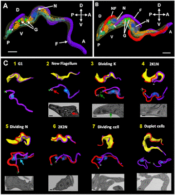
Patterns of organelle ontogeny through a cell cycle revealed by whole-cell reconstructions using 3D electron microscopy

Louise Hughes, Samantha Borrett, Katie Towers, Tobias Starborg, Sue Vaughan

J Cell Sci 2017 130: 637-647; doi: 10.1242/jcs.198887


ABSTRACT

The major mammalian bloodstream form of the African sleeping sickness parasite Trypanosoma brucei multiplies rapidly, and it is important to understand how these cells divide. Organelle inheritance involves complex spatiotemporal re-arrangements to ensure correct distribution to daughter cells. Here, serial block face scanning electron microscopy (SBF-SEM) was used to reconstruct whole individual cells at different stages of the cell cycle to give an unprecedented temporal, spatial and quantitative view of organelle division, inheritance and abscission in a eukaryotic cell. Extensive mitochondrial branching occurred only along the ventral surface of the parasite, but the mitochondria returned to a tubular form during cytokinesis. Fission of the mitochondrion occurred within the cytoplasmic bridge during the final stage of cell division, correlating with cell abscission. The nuclei were located underneath each flagellum at mitosis and the mitotic spindle was located along the ventral surface, further demonstrating the asymmetric arrangement of cell cleavage in trypanosomes. Finally, measurements demonstrated that multiple Golgi bodies were accurately positioned along the flagellum attachment zone, suggesting a mechanism for determining the location of Golgi bodies along each flagellum during the cell cycle.

FREE PDF GRATIS: Journal of Cell Science

Visualização de relance da actina

Actin visualization at a glance

Michael Melak, Matthias Plessner, Robert Grosse

J Cell Sci 2017 130: 525-530; doi: 10.1242/jcs.189068


ABSTRACT

Actin functions in a multitude of cellular processes owing to its ability to polymerize into filaments, which can be further organized into higher-order structures by an array of actin-binding and regulatory proteins. Therefore, research on actin and actin-related functions relies on the visualization of actin structures without interfering with the cycles of actin polymerization and depolymerization that underlie cellular actin dynamics. In this Cell Science at a Glance and the accompanying poster, we briefly evaluate the different techniques and approaches currently applied to analyze and visualize cellular actin structures, including in the nuclear compartment. Referring to the gold standard F-actin marker phalloidin to stain actin in fixed samples and tissues, we highlight methods for visualization of actin in living cells, which mostly apply the principle of genetically fusing fluorescent proteins to different actin-binding domains, such as LifeAct, utrophin and F-tractin, as well as anti-actin-nanobody technology. In addition, the compound SiR-actin and the expression of GFP–actin are also applicable for various types of live-cell analyses. Overall, the visualization of actin within a physiological context requires a careful choice of method, as well as a tight control of the amount or the expression level of a given detection probe in order to minimize its influence on endogenous actin dynamics.

FREE PDF GRATIS: Journal of Cell Science