Metabiota - um site sobre epidemias no mundo inteiro

sexta-feira, abril 19, 2019

Resultado de imagem para Metabiota
Um site rastreador sobre epidemias no mundo inteiro.


Origem do RNA codificante a partir de sequência aleatória de RNA

quarta-feira, abril 17, 2019

Origin of Coding RNA from Random-Sequence RNA

Gaspar Banfalvi

Published Online:5 Mar 2019 https://doi.org/10.1089/dna.2018.4389


FIG. 1. Ribose, the best fitting pentose in the nucleotide structure.

Abstract

D-ribose and D-arabinose differ only by the steric orientation of their C2-OH groups. The initial reactions and emergence of RNA depended on the position, reactivity, and flexibility of the C2-OH moiety in the ribose molecule. The steric relationship of the C2- and C3-OH groups favored the selection of ribose, ribonucleotide, and RNA synthesis and excluded the possibility of xenonucleic acid-based life on Earth. This brief review provides a hypothesis based on the absence of nucleotides and enzymes under prebiotic conditions and on the polymerization of ribose 5-phosphate units leading to the polarized formation of the ribose-phosphate backbone. The strong covalent bond formation in the sugar-phosphate backbone was followed by the somewhat less reactive interaction between ribose and nucleobase and supplemented by even weaker hydrogen-bonded and stacking interactions. This hypothesis proposes a scheme how prebiotic random-sequence RNA was formed under abiotic conditions and hydrolyzed to oligomers and nucleotides. The term random-sequence prebiotic RNA refers to nucleobases attached randomly to the ribose-phosphate backbone and not to cellular RNA sequences as proteins and cells did not probably exist at the time of abiotic RNA formation. It is hypothesized that RNA generated under abiotic conditions containing random nucleobases was hydrolyzed to nucleotides that served as a pool for the selected synthesis of genetic RNA.

FREE PDF GRATIS: DNA and Cell Biology

Nova hipótese da origem da vida: concentrações de óxido de nitrogênio em águas naturais na Terra Primeva

sábado, abril 13, 2019

Geochemistry, Geophysics, Geosystems

Nitrogen Oxide Concentrations in Natural Waters on Early Earth

Sukrit Ranjan  Zoe R. Todd  Paul B. Rimmer  Dimitar D. Sasselov  Andrew R. Babbin

First published: 12 April 2019 https://doi.org/10.1029/2018GC008082

This article has been accepted for publication and undergone full peer review but has not been through the copyediting, typesetting, pagination and proofreading process, which may lead to differences between this version and the Version of Record. Please cite this article as doi: 10.1029/2018gc008082

Resultado de imagem para early earth images


Abstract


A key challenge in origins‐of‐life studies is estimating the abundances of species relevant to the chemical pathways proposed to have contributed to the emergence of life on early Earth. Dissolved nitrogen oxide anions (NOurn:x-wiley:ggge:media:ggge21866:ggge21866-math-0001), in particular nitrate (NOurn:x-wiley:ggge:media:ggge21866:ggge21866-math-0002) and nitrite (NOurn:x-wiley:ggge:media:ggge21866:ggge21866-math-0003), have been invoked in diverse origins‐of‐life chemistry, from the oligomerization of RNA to the emergence of protometabolism. Recent work has calculated the supply of NOurn:x-wiley:ggge:media:ggge21866:ggge21866-math-0018 from the prebiotic atmosphere to the ocean, and reported steady‐state [NOurn:x-wiley:ggge:media:ggge21866:ggge21866-math-0005] to be high across all plausible parameter space. These findings rest on the assumption that NOurn:x-wiley:ggge:media:ggge21866:ggge21866-math-0004 is stable in natural waters unless processed at a hydrothermal vent. Here, we show that NOurn:x-wiley:ggge:media:ggge21866:ggge21866-math-0006 is unstable in the reducing environment of early Earth. Sinks due to UV photolysis and reactions with reduced iron (urn:x-wiley:ggge:media:ggge21866:ggge21866-math-0019) suppress [NOurn:x-wiley:ggge:media:ggge21866:ggge21866-math-0007] by several orders of magnitude relative to past predictions. For pH= 6.5 ‐ 8 and T=0‐50°C, we find that it is most probable that [NOurn:x-wiley:ggge:media:ggge21866:ggge21866-math-0008]<1 alt="urn:x-wiley:ggge:media:ggge21866:ggge21866-math-0020" class="section_image" img="" nbsp="" src="https://wol-prod-cdn.literatumonline.com/cms/attachment/2a0116a6-29d1-44f7-b669-6d2697d1616e/ggge21866-math-0020.png" style="border-style: none; box-sizing: border-box; max-width: 100%; vertical-align: middle;">M in the prebiotic ocean. On the other hand, prebiotic ponds with favorable drainage characteristics may have sustained [NOurn:x-wiley:ggge:media:ggge21866:ggge21866-math-0009]≥1μM. As on modern Earth, most NOurn:x-wiley:ggge:media:ggge21866:ggge21866-math-0010 on prebiotic Earth should have been present as NO urn:x-wiley:ggge:media:ggge21866:ggge21866-math-0011, due to its much greater stability. These findings inform the kind of prebiotic chemistries that would have been possible on early Earth. We discuss the implications for proposed prebiotic chemistries, and highlight the need for further studies of NOurn:x-wiley:ggge:media:ggge21866:ggge21866-math-0012 kinetics to reduce the considerable uncertainties in predicting [NOurn:x-wiley:ggge:media:ggge21866:ggge21866-math-0013] on early Earth.

Pele de dinossauro do Cretáceo perfeitamente preservada encontrada na Coreia

quinta-feira, abril 11, 2019

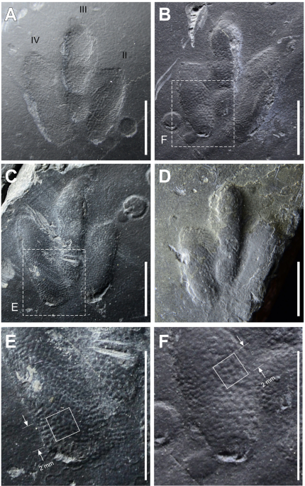
Exquisitely-preserved, high-definition skin traces in diminutive theropod tracks from the Cretaceous of Korea

Kyung Soo Kim, Martin G. Lockley, Jong Deock Lim & Lida Xing 

Scientific Reports volume 9, Article number: 2039 (2019)

Figure 4
Fig. 4

Abstract

Small theropod tracks, ichnogenus Minisauripus, from the Jinju Formation (Cretaceous) of Korea reveal exquisitely preserved skin texture impressions. This is the first report for any dinosaur of skin traces that cover entire footprints, and every footprint in a trackway. Special sedimentological conditions allowed footprint registration without smearing of skin texture patterns which consist of densely-packed, reticulate arrays of small (<0 .5="" also="" and="" as="" avian="" birds="" both="" casts="" china="" cretaceous="" different="" essentially="" foot="" for="" from="" had="" impressions="" is="" latter="" lower="" mm="" morphologies.="" nbsp="" of="" oldest="" polygons="" preserved="" quite="" replicas.="" report="" reported="" resembles="" skin="" span="" texture="" that="" the="" theropods="" this="" two="" which="">Minisauripus from Korea predating five reports from the Haman Formation of inferred Albian age. Minisauripus is now known from six Korean and three Chinese localities, all from the Lower Cretaceous. This gives a total sample of ~ 95 tracks representing ~ 54 trackways. With 80% of tracks <3 .0="" cm="" long="" nbsp="" span="">Minisauripus is pivotal in debates over whether small tracks represent small species, as the database suggests, or juveniles of large species. 

Acknowledgements

We thank the School of Biological Sciences, the University of Queensland, Brisbane for help with statistical analyses done in Supplementary Information.

Author information

Affiliations

Department of Science Education, Chinju National University of Education, 3 Jinnyangho-ro 369beon-gil, Jinju-si, Gyeongnam, 52673, Korea
Kyung Soo Kim

Dinosaur Trackers Research Group, University of Colorado Denver, P.O. Box 173364, Denver, CO, 80217, USA
Martin G. Lockley

Cultural Heritage Administration, Government Complex-Daejeon, 189, Cheongsa-ro, Seo-gu, Daejon, 35208, Korea
Jong Deock Lim

School of the Earth Sciences and Resources, China University of Geosciences, Beijing, 100083, China
Lida Xing

Contributions

K.-S.K. found, collected and photographed specimens K.-S.K., M.G.L. and J.-D.L. examined field site, measured specimens and prepared manuscript and figures. L.X. examined comparative material and helped with bibliographic research and database organization.

Competing Interests

The authors declare no competing interests.

Corresponding author

Correspondence to Martin G. Lockley.

Rights and permissions

Creative Commons BY

Open Access This article is licensed under a Creative Commons Attribution 4.0 International License, which permits use, sharing, adaptation, distribution and reproduction in any medium or format, as long as you give appropriate credit to the original author(s) and the source, provide a link to the Creative Commons license, and indicate if changes were made. The images or other third party material in this article are included in the article’s Creative Commons license, unless indicated otherwise in a credit line to the material. If material is not included in the article’s Creative Commons license and your intended use is not permitted by statutory regulation or exceeds the permitted use, you will need to obtain permission directly from the copyright holder. To view a copy of this license, visit http://creativecommons.org/licenses/by/4.0/.

About this article

Publication history

Received 28 September 2018 Accepted 03 January 2019

Published 14 February 2019

DOI


Subjects Palaeontology Solid Earth sciences

FREE PDF GRATIS: Scientific Reports Sup. Info.

Insights estruturais sobre características únicas da reciclagem de ribossomos mitocondriais humanos

quarta-feira, abril 10, 2019

Structural insights into unique features of the human mitochondrial ribosome recycling

Ravi K. Koripella, Manjuli R. Sharma, Paul Risteff, Pooja Keshavan, and Rajendra K. Agrawal

PNAS published ahead of print April 8, 2019 


Edited by Yale E. Goldman, Pennsylvania Muscle Institute, University of Pennsylvania, Philadelphia, PA, and approved March 15, 2019 (received for review September 11, 2018)

Image result for human mitochondrial ribosome recycling

Significance

The human mitochondrial ribosome (mitoribosome) recycling factor (RRFmt) is known to play essential roles in mitochondrial physiology, including protein synthesis, and it has been implicated in human genetic diseases. The RRFmt is among the few protein molecules that carry their N-terminal signal peptide sequence into the mitochondrial matrix that is required for RRFmt’s interaction with the mitoribosome. In this study, we present a cryo-electron microscopic structure of the human mitoribosome in complex with the RRFmt. The structure reveals hitherto unknown features of RRFmt and its interactions with the functionally important regions of the ribosomal RNA that constitute the peptidyl-transferase center and that are linked to the GTPase-associated center of the mitoribosome, shedding light on the mechanism of ribosome recycling in mitochondria.

Abstract

Mammalian mitochondrial ribosomes (mitoribosomes) are responsible for synthesizing proteins that are essential for oxidative phosphorylation (ATP generation). Despite their common ancestry with bacteria, the composition and structure of the human mitoribosome and its translational factors are significantly different from those of their bacterial counterparts. The mammalian mitoribosome recycling factor (RRFmt) carries a mito-specific N terminus extension (NTE), which is necessary for the function of RRFmt. Here we present a 3.9-Å resolution cryo-electron microscopic (cryo-EM) structure of the human 55S mitoribosome-RRFmt complex, which reveals α-helix and loop structures for the NTE that makes multiple mito-specific interactions with functionally critical regions of the mitoribosome. These include ribosomal RNA segments that constitute the peptidyl transferase center (PTC) and those that connect PTC with the GTPase-associated center and with mitoribosomal proteins L16 and L27. Our structure reveals the presence of a tRNA in the pe/E position and a rotation of the small mitoribosomal subunit on RRFmt binding. In addition, we observe an interaction between the pe/E tRNA and a mito-specific protein, mL64. These findings help understand the unique features of mitoribosome recycling.

human mitochondrial RRFmito-specific sequence55S–RRFmt complexcryo-EM structuremito-specific interactions

Footnotes

↵1Present address: Charles River Laboratories Inc., Durham, NC 27703.

↵2To whom correspondence should be addressed. Email: Rajendra.Agrawal@health.ny.gov.
Author contributions: R.K.A. designed research; R.K.K., M.R.S., P.R., and P.K. performed research; R.K.K., M.R.S., and R.K.A. analyzed data; and R.K.K., M.R.S., and R.K.A. wrote the paper.

The authors declare no conflict of interest.

This article is a PNAS Direct Submission.

Data deposition: The cryo-EM maps and atomic coordinates have been deposited in the Electron Microscopy and PDB Data Bank (www.wwpdb.org) under accession codes EMD-0514 and PDB ID 6NU2, respectively, for the RRFmt-bound 55S mitoribosome (Complex I) and EMD-0515 and PDB ID 6NU3, respectively, for the unbound 55S mitoribosome (Complex II).

FREE PDF GRATIS: PNAS Sup. Info.

Copyright © 2019 the Author(s). Published by PNAS.

Pesquisadores repensam a ancestralidade de células complexas

Researchers Rethink the Ancestry of Complex Cells
New studies revise ideas about the symbiosis that gave mitochondria to cells and about whether the last common ancestor of all eukaryotes was one cell or many.

Research into the origins of the complex cells called eukaryotes typically tries to trace lineages of the organisms back to a single ancestral cell. But now scientists are considering whether that common “ancestor” might really be an entire population of diverse cells.

DVDP for Quanta Magazine

Our planet formed a little over 4.5 billion years ago, and if the most recent estimates are correct, it wasn’t long before life arose. Not much is known about how that happened because it’s maddeningly difficult to investigate. It’s also proved tough to study what happened next, during the first billions of years of evolution that followed, when the main domains of life emerged.

A particularly vexing mystery is the rise of the eukaryotes, cells with well-defined internal compartments, or organelles, which are present only in animals, plants, fungi and some microbes like protists — our evolutionary kin. The earliest eukaryotes left no clear fossils as clues, so researchers are forced to deduce what they were like by comparing the structural and molecular details of later ones and inferring their evolutionary relationships.

Right now is “an incredibly exciting time” for such research, said Michelle Leger, a postdoctoral fellow at the Institute of Evolutionary Biology in Barcelona, Spain. With modern genetic sequencing technologies, scientists can read the entire genomes of diverse life forms, and as microbial life is revealed in ever-increasing detail, new species and other taxonomic groups are coming to light. With that wealth of data, researchers are tracing lineages of organisms backward through time. “We’re trying to approach the problem from so many sides,” she said. “That’s pushing us closer to the first eukaryotes.”


This paramecium, a single-cell protozoan microbe, has a nucleus, mitochondria and other organelles that are hallmarks of eukaryotic cells.

Michael Abbey/Science Source

And those first eukaryotes may depart significantly from what most scientists expected, if some recent findings are any indication. Earlier this month, one team presented evidence that a signature event in eukaryote evolution — the development of the organelles called mitochondria — might have unfolded quite differently than was theorized. Meanwhile, other researchers have suggested that the earliest “ancestor” of all eukaryotes might not have been a single cell at all, but rather a mixed population of cells that avidly swapped DNA. The difference is subtle, but it might be important for understanding the evolution and diversity of the eukaryotes we see today.

The Ancestral Eukaryotes

The very first cells — the first life forms on this planet — were prokaryotes, but they were not all alike. Even early on, two very distinct lineages emerged, the archaea and the bacteria. The archaea might have been the first to thrive because even now they can survive in extreme environments like hot vents and super-saline pools. But it’s also possible that archaea and bacteria split from the first cells at the same time and began to diversify independently from the start. Figuring out definitively when and how the split occurred is probably impossible given how much time has passed; fossil evidence is nonexistent, and organisms from both branches have swapped genes extensively through horizontal gene transfer (as opposed to the “vertical” transfer of genes down through generations), which complicates analyses of their genomic history.

What we do know is that the story of eukaryotes began when some rogue archaeal cell split from the rest and founded what was long considered an entirely new domain of life. “First and fundamentally we are a very strange kind of archaea,” said Maureen O’Malley, a philosopher of biology affiliated with the University of Bordeaux and the University of Sydney.
...

Read more here: Quanta Magazine

A replicação do DNA não é uniforme e nem consistente

segunda-feira, abril 08, 2019

Recycling of single-stranded DNA-binding protein by the bacterial replisome 

Lisanne M Spenkelink  Jacob S Lewis  Slobodan Jergic  Zhi-Qiang Xu  Andrew Robinson Nicholas E Dixon  Antoine M van Oijen

Nucleic Acids Research, gkz090, https://doi.org/10.1093/nar/gkz090

Published: 15 February 2019  

Article history
Received: 18 September 2018 Revision Received: 30 January 2019
Accepted: 09 February 2019


Abstract

Single-stranded DNA-binding proteins (SSBs) support DNA replication by protecting single-stranded DNA from nucleolytic attack, preventing intra-strand pairing events and playing many other regulatory roles within the replisome. Recent developments in single-molecule approaches have led to a revised picture of the replisome that is much more complex in how it retains or recycles protein components. Here, we visualize how an in vitro reconstituted Escherichia coli replisome recruits SSB by relying on two different molecular mechanisms. Not only does it recruit new SSB molecules from solution to coat newly formed single-stranded DNA on the lagging strand, but it also internally recycles SSB from one Okazaki fragment to the next. We show that this internal transfer mechanism is balanced against recruitment from solution in a manner that is concentration dependent. By visualizing SSB dynamics in live cells, we show that both internal transfer and external exchange mechanisms are physiologically relevant.

A abordagem científica para evolução: o que eles não lhe ensinaram em Biologia

domingo, abril 07, 2019

The Scientific Approach to Evolution: What They Didn't Teach You in Biology 

Paperback – September 14, 2016

by Rob Stadler (Author)

For more than 150 years, continuous debate has swirled around the topic of evolution. From Darwin to Dawkins, extensive scientific evidence has been presented for evolution, yet almost half of contemporary society still isn’t convinced. The Scientific Approach to Evolution provides a rational new perspective on this debate. Scientific evidence is not all created equally. Some forms of evidence provide only low confidence, while other forms of evidence provide high confidence. Rob Stadler describes a compelling approach to determine the level of confidence and applies it to the commonly cited evidence for evolution. When high-confidence evidence is appropriately prioritized over low-confidence evidence, the result is a profound new view of evolution—one that they did not teach you in biology.

About the Author

Rob Stadler received a BS in biomedical engineering from Case Western Reserve University, an MS in electrical engineering from MIT, and a PhD in medical engineering from the Harvard/MIT Division of Health Sciences and Technology. With 19 years of experience as a scientist in the medical device industry, he has authored 17 peer-reviewed manuscripts, has obtained more than 100 US patents, and his research and innovation have contributed to medical devices that are implanted in over 1 million patients worldwide.

Source/Fonte: Amazon

Dollo, quem foi que disse que a evolução era irreversível???

sábado, abril 06, 2019

Journal of Evolutionary Biology Volume 0, Issue 0

Opposite responses to selection and where to find them

David N. Fisher  Jonathan N. Pruitt

First published: 26 February 2019


Source/Fonte: ClipartPanda

Abstract

We generally expect traits to evolve in the same direction as selection. However, many organisms possess traits that appear to be costly for individuals, while plant and animal breeding experiments reveal that selection may lead to no response or even negative responses to selection. We formalize both of these instances as cases of “opposite responses to selection.” Using quantitative genetic models for the response to selection, we outline when opposite responses to selection should be expected. These typically occur when social selection opposes direct selection, when individuals interact with others less related to them than a random member of the population, and if the genetic covariance between direct and indirect effects is negative. We discuss the likelihood of each of these occurring in nature and therefore summarize how frequent opposite responses to selection are likely to be. This links several evolutionary phenomena within a single framework.

Inovação Evolutiva Induzida por Estresse: Um Mecanismo para a Origem dos Tipos de Células

Stress‐Induced Evolutionary Innovation: A Mechanism for the Origin of Cell Types

Prof. Günter P. Wagner  Dr. Eric M. Erkenbrack  Prof. Alan C. Love

First published: 28 March 2019


Fig. 1: Evolutionary history of viviparity and the DSC in mammals. 

Abstract

Understanding the evolutionary role of environmentally induced phenotypic variation (i.e., plasticity) is an important issue in developmental evolution. A major physiological response to environmental change is cellular stress, which is counteracted by generic stress reactions detoxifying the cell. A model, stress‐induced evolutionary innovation (SIEI), whereby ancestral stress reactions and their corresponding pathways can be transformed into novel structural components of body plans, such as new cell types, is described. Previous findings suggest that the cell differentiation cascade of a cell type critical to pregnancy in humans, the decidual stromal cell, evolved from a cellular stress reaction. It is hypothesized that the stress reaction in these cells was elicited ancestrally via inflammation caused by embryo attachment. The present study proposes that SIEI is a distinct form of plasticity‐based evolutionary change leading to the origin of novel structures rather than adaptive transformation of pre‐existing characters.

FREE PDF GRATIS: BioEssays

A estrutura emergente da Síntese Evolutiva Ampliada/Estendida: onde se encaixa a Evo-Devo?

sexta-feira, abril 05, 2019

Theory in Biosciences

November 2018, Volume 137, Issue 2, pp 169–184 | Cite as

The emerging structure of the Extended Evolutionary Synthesis: where does Evo-Devo fit in?

Alejandro Fábregas-Tejeda, Francisco Vergara-Silva

First Online: 21 August 2018


Abstract

The Extended Evolutionary Synthesis (EES) debate is gaining ground in contemporary evolutionary biology. In parallel, a number of philosophical standpoints have emerged in an attempt to clarify what exactly is represented by the EES. For Massimo Pigliucci, we are in the wake of the newest instantiation of a persisting Kuhnian paradigm; in contrast, Telmo Pievani has contended that the transition to an EES could be best represented as a progressive reformation of a prior Lakatosian scientific research program, with the extension of its Neo-Darwinian core and the addition of a brand-new protective belt of assumptions and auxiliary hypotheses. Here, we argue that those philosophical vantage points are not the only ways to interpret what current proposals to ‘extend’ the Modern Synthesis-derived ‘standard evolutionary theory’ (SET) entail in terms of theoretical change in evolutionary biology. We specifically propose the image of the emergent EES as a vast network of models and interweaved representations that, instantiated in diverse practices, are connected and related in multiple ways. Under that assumption, the EES could be articulated around a paraconsistent network of evolutionary theories (including some elements of the SET), as well as models, practices and representation systems of contemporary evolutionary biology, with edges and nodes that change their position and centrality as a consequence of the co-construction and stabilization of facts and historical discussions revolving around the epistemic goals of this area of the life sciences. We then critically examine the purported structure of the EES—published by Laland and collaborators in 2015—in light of our own network-based proposal. Finally, we consider which epistemic units of Evo-Devo are present or still missing from the EES, in preparation for further analyses of the topic of explanatory integration in this conceptual framework.

Keywords

Extended Evolutionary Synthesis Evolutionary biology Paradigm Scientific research program Epistemic units Evo-Devo 

Notes

Acknowledgements

The authors acknowledge the advice and assistance of Fátima Sofía Ávila-Cascajares during the conception of Fig. 1, and thank Casandra Lizbeth Méndez-Martínez for her aid in the design of Fig. 1. Diana Martínez Almaguer and Julio César Montero Rojas (Graphic Design Unit, Instituto de Biología, UNAM) assisted in the design of all figures of the article. We also thank Mario Casanueva for discussions and his critical remarks about the structure of the EES. The comments and feedback received from Alan Love and Kevin Laland during the presentation of this work at the 2017 Meeting of the International Society for History, Philosophy and Social Studies of Biology (ISHPSSB) in São Paulo, Brazil, were valuable for the preparation of revised versions of this work; at the same meeting, we also benefited from conversations with Eva Jablonka, Marion Lamb and Jan Baedke about the sociological, epistemological and political dimensions involved in the EES debate. AFT is indebted to participants in the poster session of the Sixth Meeting of the European Society for Evolutionary Developmental Biology (Uppsala, July 2016), especially to Mark Jonas. AFT also thanks Francesco Suman for comments on an earlier version of this manuscript and discussions on the structure of the EES and the pluralist landscape of contemporary evolutionism (Washington D.C., September 2016). FVS acknowledges facilities provided by libraries at the Instituto de Biología, UNAM and other academic institutions in Mexico City, Uppsala and London during his long-term research projects on the historiography, epistemology and sociology of biology. Finally, we thank an anonymous reviewer for critical comments which substantially improved the original manuscript. The authors declare that they have no conflict of interest, and that they did not receive any specific funding during the research and writing of this work.

Darwin, mutações não aleatórias em micróbios "comprometem" a síntese moderna

quinta-feira, abril 04, 2019

What is mutation? A chapter in the series: How microbes “jeopardize” the modern synthesis

Devon M. Fitzgerald, Susan M. Rosenberg 


Abstract

Mutations drive evolution and were assumed to occur by chance: constantly, gradually, roughly uniformly in genomes, and without regard to environmental inputs, but this view is being revised by discoveries of molecular mechanisms of mutation in bacteria, now translated across the tree of life. These mechanisms reveal a picture of highly regulated mutagenesis, up-regulated temporally by stress responses and activated when cells/organisms are maladapted to their environments—when stressed—potentially accelerating adaptation. Mutation is also nonrandom in genomic space, with multiple simultaneous mutations falling in local clusters, which may allow concerted evolution—the multiple changes needed to adapt protein functions and protein machines encoded by linked genes. Molecular mechanisms of stress-inducible mutation change ideas about evolution and suggest different ways to model and address cancer development, infectious disease, and evolution generally.

Citation: Fitzgerald DM, Rosenberg SM (2019) What is mutation? A chapter in the series: How microbes “jeopardize” the modern synthesis. PLoS Genet 15(4): e1007995. https://doi.org/10.1371/journal.pgen.1007995

Editor: W. Ford Doolittle, Dalhousie University, CANADA

Published: April 1, 2019

Copyright: © 2019 Fitzgerald, Rosenberg. This is an open access article distributed under the terms of the Creative Commons Attribution License, which permits unrestricted use, distribution, and reproduction in any medium, provided the original author and source are credited.

Funding: This work was supported by the American Cancer Society Postdoctoral Fellowship 132206-PF-18-035-01-DMC (DMF) and NIH grant R35-GM122598. The funders had no role in the preparation of the article.

Competing interests: The authors have declared that no competing interests exist.

FREE PDF GRATIS: PLoS Genetics

Reescrita de síntese química de um genoma bacteriano para alcançar flexibilidade de design e funcionalidade biológica

terça-feira, abril 02, 2019

Chemical synthesis rewriting of a bacterial genome to achieve design flexibility and biological functionality

Jonathan E. Venetz, Luca Del Medico, Alexander Wölfle, Philipp Schächle, Yves Bucher, Donat Appert, Flavia Tschan, Carlos E. Flores-Tinoco, Mariëlle van Kooten, Rym Guennoun, Samuel Deutsch, Matthias Christen, and Beat Christen

PNAS published ahead of print April 1, 2019 


Edited by David Baker, University of Washington, Seattle, WA, and approved March 6, 2019 (received for review October 29, 2018)


Sequence design flexibility within rewritten C. eth-2.0 genes. 
Source/Fonte: PNAS

Significance

The fundamental biological functions of a living cell are stored within the DNA sequence of its genome. Classical genetic approaches dissect the functioning of biological systems by analyzing individual genes, yet uncovering the essential gene set of an organism has remained very challenging. It is argued that the rewriting of entire genomes through the process of chemical synthesis provides a powerful and complementary research concept to understand how essential functions are programed into genomes.

Abstract

Understanding how to program biological functions into artificial DNA sequences remains a key challenge in synthetic genomics. Here, we report the chemical synthesis and testing of Caulobacter ethensis-2.0 (C. eth-2.0), a rewritten bacterial genome composed of the most fundamental functions of a bacterial cell. We rebuilt the essential genome of Caulobacter crescentus through the process of chemical synthesis rewriting and studied the genetic information content at the level of its essential genes. Within the 785,701-bp genome, we used sequence rewriting to reduce the number of encoded genetic features from 6,290 to 799. Overall, we introduced 133,313 base substitutions, resulting in the rewriting of 123,562 codons. We tested the biological functionality of the genome design in C. crescentus by transposon mutagenesis. Our analysis revealed that 432 essential genes of C. eth-2.0, corresponding to 81.5% of the design, are equal in functionality to natural genes. These findings suggest that neither changing mRNA structure nor changing the codon context have significant influence on biological functionality of synthetic genomes. Discovery of 98 genes that lost their function identified essential genes with incorrect annotation, including a limited set of 27 genes where we uncovered noncoding control features embedded within protein-coding sequences. In sum, our results highlight the promise of chemical synthesis rewriting to decode fundamental genome functions and its utility toward the design of improved organisms for industrial purposes and health benefits.

Caulobacter crescentus chemical genome synthesis genome rewriting synonymous recoding de novo DNA synthesis

Footnotes

↵1To whom correspondence may be addressed. Email: matthias.christen@imsb.biol.ethz.ch or beat.christen@imsb.biol.ethz.ch.

Author contributions: M.C. and B.C. designed research; J.E.V., L.D.M., A.W., P.S., Y.B., D.A., F.T., C.E.F.-T., M.v.K., R.G., and S.D. performed research; J.E.V., L.D.M., M.C., and B.C. analyzed data; and J.E.V., M.C., and B.C. wrote the paper.

Conflict of interest statement: Eidgenössische Technische Hochschule holds a patent application (WO2017085249A1) with M.C. and B.C. as inventors that covers functional testing of synthetic genomes. M.C. and B.C. hold shares from Gigabases Switzerland AG.

This article is a PNAS Direct Submission.

Data deposition: The sequence of the C. eth-2.0 genome reported in this paper has been deposited in the National Center for Biotechnology Information database (GenBank accession no. CP035535).

FREE PDF GRATIS: PNAS Supporting Information

Copyright © 2019 the Author(s). Published by PNAS.

This open access article is distributed under Creative Commons Attribution-NonCommercial-NoDerivatives License 4.0 (CC BY-NC-ND).

A ecologia e evolução dos sistemas imune adaptativos microbianos CRISPR-Cas

The ecology and evolution of microbial CRISPR-Cas adaptive immune systems

Edze R. Westra, Stineke van Houte, Sylvain Gandon and Rachel Whitaker

Published:25 March 2019Article ID:20190101


1. Introduction

Over the past decade, the field of CRISPR-Cas research has received a lot of attention from the scientific community. While initially, this mostly concerned microbiologists who were fascinated by the discovery that some bacteria encode RNA-guided adaptive immune systems, this rapidly spread to other scientific disciplines following the development of groundbreaking molecular biology tools [1] and, more recently, to the public domain where the societal and ethical implications and legislation surrounding CRISPR applications are being debated. Some of the potential CRISPR applications that are currently being explored in the laboratory would involve the release of CRISPR genes into confined or open environments—for example, when CRISPR would be used to protect focal bacterial species against phage infections, when it is applied to suppress the spread of antimicrobial resistance or to control vectors of disease [2– 4]. One component of the debate surrounding the societal impacts of these applications entails an assessment of the potential risks associated with these strategies (e.g. [5– 7]), which requires an understanding of how CRISPR-Cas behaves in an ecological context. In this special issue, we explore this question by examining the evolutionary history of CRISPR-Cas immune systems, where they occur naturally, when they evolve and how this impacts the spread and evolution of other DNA elements. Finally, we return to the question how CRISPR-Cas may be exploited in an ecological context for the benefit of human health, and the ethical challenges that are associated with this.

FREE PDF GRATIS: Phil Trans R Soc B