Patterns of organelle ontogeny through a cell cycle revealed by whole-cell reconstructions using 3D electron microscopy
Louise Hughes, Samantha Borrett, Katie Towers, Tobias Starborg, Sue Vaughan
J Cell Sci 2017 130: 637-647; doi: 10.1242/jcs.198887
ABSTRACT
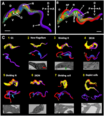
The major mammalian bloodstream form of the African sleeping sickness parasite Trypanosoma brucei multiplies rapidly, and it is important to understand how these cells divide. Organelle inheritance involves complex spatiotemporal re-arrangements to ensure correct distribution to daughter cells. Here, serial block face scanning electron microscopy (SBF-SEM) was used to reconstruct whole individual cells at different stages of the cell cycle to give an unprecedented temporal, spatial and quantitative view of organelle division, inheritance and abscission in a eukaryotic cell. Extensive mitochondrial branching occurred only along the ventral surface of the parasite, but the mitochondria returned to a tubular form during cytokinesis. Fission of the mitochondrion occurred within the cytoplasmic bridge during the final stage of cell division, correlating with cell abscission. The nuclei were located underneath each flagellum at mitosis and the mitotic spindle was located along the ventral surface, further demonstrating the asymmetric arrangement of cell cleavage in trypanosomes. Finally, measurements demonstrated that multiple Golgi bodies were accurately positioned along the flagellum attachment zone, suggesting a mechanism for determining the location of Golgi bodies along each flagellum during the cell cycle.
FREE PDF GRATIS: Journal of Cell Science


